
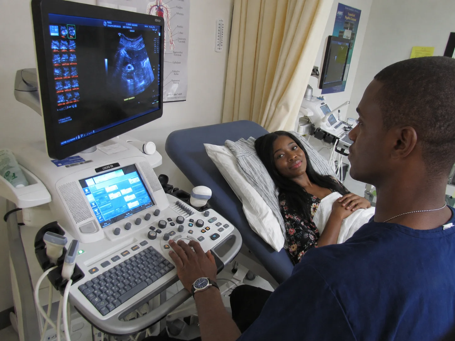
BUYER’S GUIDE / ULTRASOUND
Choosing an ultrasound system: applications, probes and total cost
A practical framework to link the clinical programme, transducers, imaging functions, workflow and maintenance.
The console alone does not define the right configuration.
An ultrasound system becomes truly useful when its probes, software, ergonomics and support match the examinations performed. Two systems in a comparable range can produce very different results depending on the patient population, the operator, the installed options and the quality of maintenance.
Start with the examinations, the patients and the operators
Describe the daily volume, the specialties, the level of urgency, the patient profiles and the users’ experience. A general radiology department, a maternity unit, an echocardiography laboratory and an emergency unit do not have the same probes, nor the same requirements for measurement, mobility and archiving.
Sort functions into three categories: essential at opening, useful in the medium term, and optional. This hierarchy prevents overpaying for rarely used licences while protecting the upgrades that are genuinely planned.
Abdomen & pelvis
Liver, biliary tract, kidneys, spleen, bladder and pelvis. Penetration, tissue harmonic imaging, Doppler and a suitable convex probe are the priorities.
Obstetrics & gynaecology
Dating, growth, foetal morphology and pelvic examination. Depending on the programme: convex, endovaginal and 3D/4D volume probes.
Cardiology
Ventricular function, valves, haemodynamics and follow-up. This requires a phased array probe, M-mode, and colour, pulsed and often continuous-wave Doppler.
Vascular
Arteries, veins, stenoses, thrombosis and vascular access. A linear probe, spectral Doppler and sensitivity to low flow are decisive.
Breast, thyroid & small parts
Superficial structures requiring high frequency, excellent contrast resolution and, depending on use, elastography.
Musculoskeletal
Tendons, muscles, joints, nerves and guided procedures. Superficial resolution, frame rate and an ergonomic linear probe matter greatly.
Paediatrics & neonatology
Higher frequencies and small-footprint probes make it easier to examine newborns, children and narrow anatomical windows.
Emergency, critical care & POCUS
Focused assessment of the heart, lungs, abdomen and volume status, plus procedural guidance. Fast start-up, battery life and simple cleaning are essential.
Choose mobility without sacrificing usability
Examination comfort, large screen, several active ports and a wide choice of options. Suited to busy departments and complex examinations.
A good compromise for travel, clinics, the operating theatre and multi-site facilities. Check battery life, durability and connectivity.
Quick access at the bedside and in the field. It often complements a fleet but does not automatically replace a full diagnostic platform.
Cardiac, women’s health or another specialty: the interface, calculations, reports and probes can be optimised for a specific care pathway.
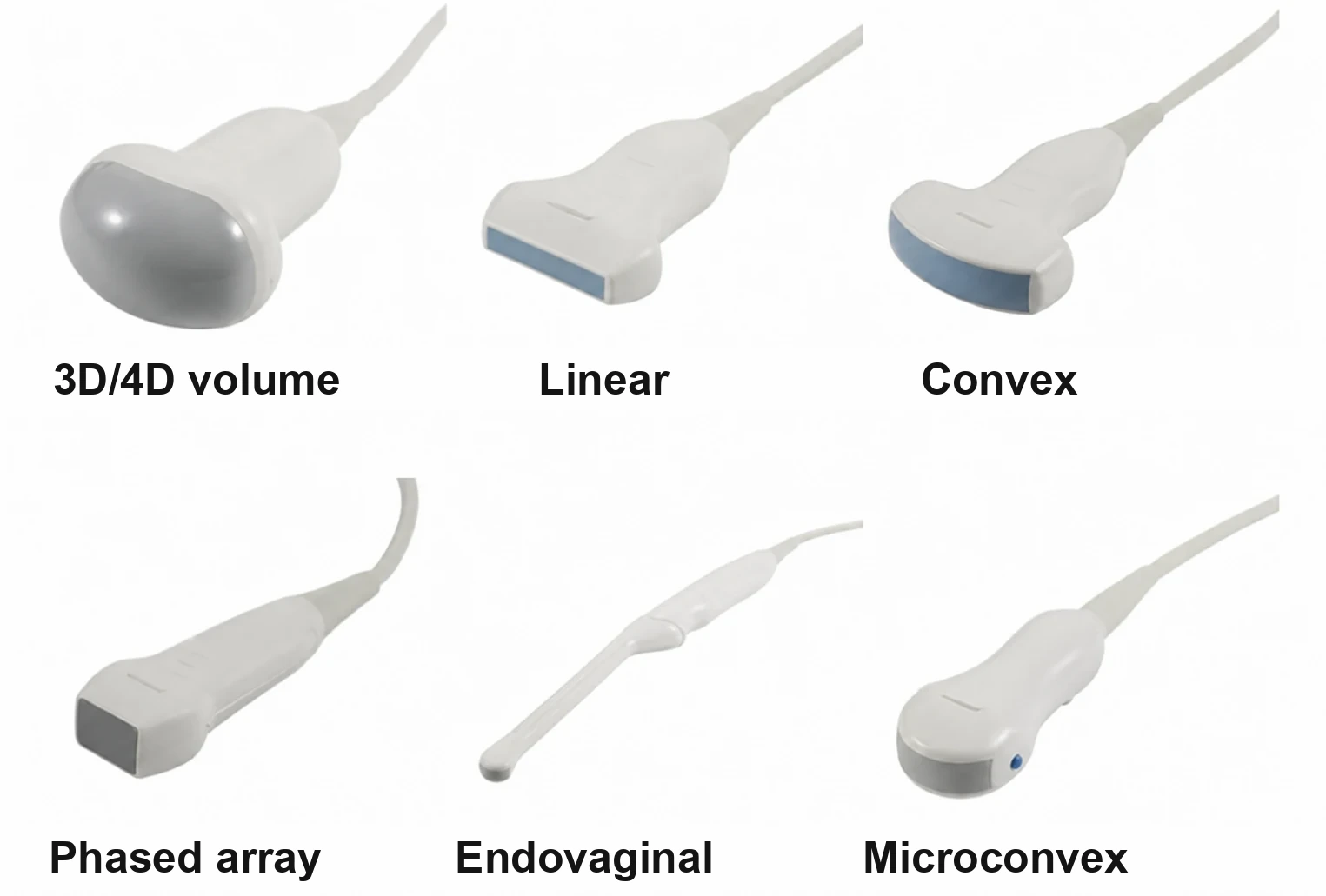
The probe is a central part of performance
A higher frequency generally improves the resolution of superficial structures, whereas a lower frequency favours penetration. The choice also depends on footprint, field of view, anatomical access, ergonomics and supported modes.
Good penetration and a wide field at depth.
High superficial resolution; penetration decreases as frequency increases.
Small footprint useful between the ribs and good access to deep structures.
Close to the target; requires high-level disinfection and suitable covers.
Combines a small footprint, a convex field and versatility.
Volume acquisition; check software, storage and reconstruction time.
Specialised, costly and fragile probe; strict reprocessing and traceability.
Continuous-wave Doppler without a 2D image on some systems; useful for high velocities.
Ask for the exact references, their compatibility with the system, the available modes, the warranty, the replacement cost and whether they can be tested on your clinical cases.
Turn options into clinical requirements
B-mode, M-mode, tissue harmonic imaging, zoom, cine, measurements and automatic optimisation form the foundation of most examinations.
Colour, power, pulsed and continuous-wave Doppler meet different needs. Check sensitivity, artefacts, frame rate and ease of adjustment.
Elastography, liver quantification and contrast can enrich certain services, but require training, protocols and suitable licences.
3D/4D, automated measurements, plane recognition and assisted tools can improve reproducibility when they are integrated into the real clinical workflow.
Needle enhancement, CT/MR fusion, navigation and contrast can be decisive for biopsies, ablations and guided procedures.
DICOM worklist, PACS storage, export, report templates, traceability and cybersecurity must be specified before purchase.
Compare several manufacturers on the same protocol
The examples below illustrate different families of systems. They are neither a ranking nor an exclusive recommendation. The availability of probes, software and services depends on the country, the version and the configuration sold.
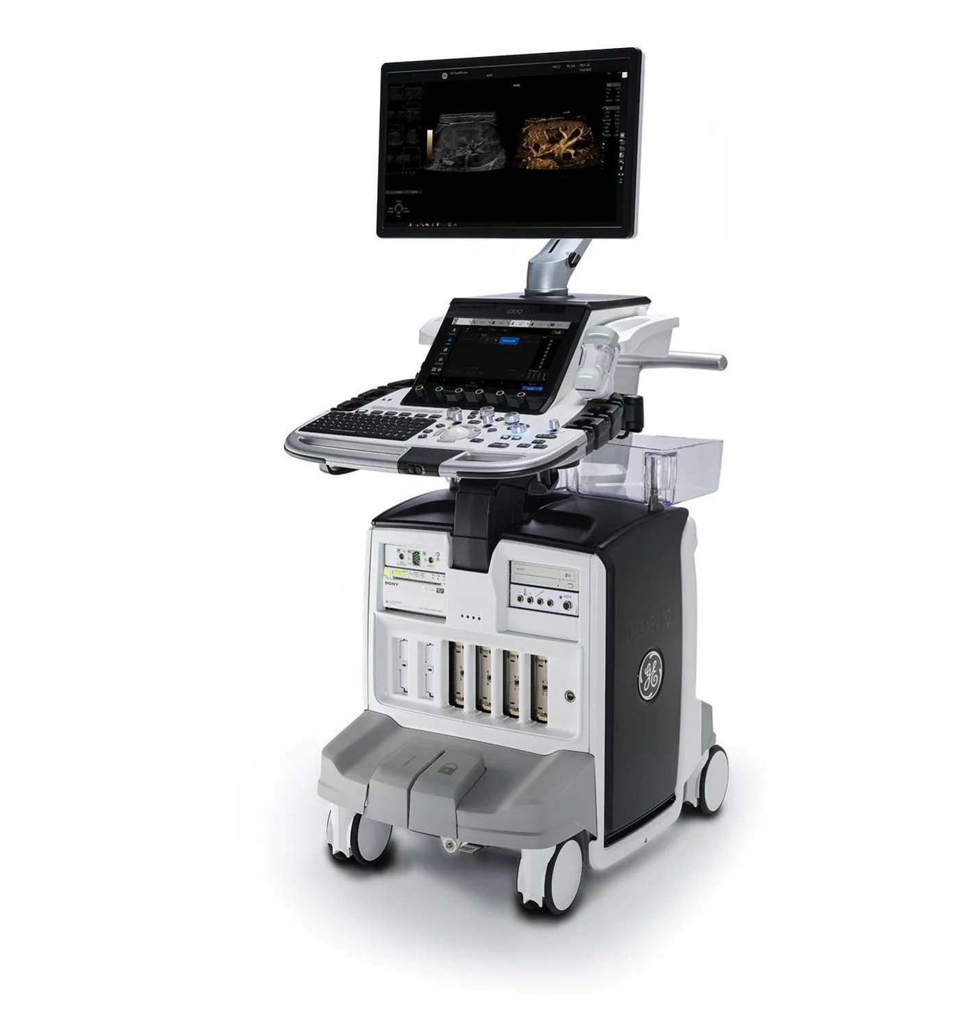
LOGIQ E10s
Example of a high-end general-purpose platform: abdominal, vascular, small-parts and interventional imaging, with advanced tools depending on configuration.
View manufacturer’s overview ↗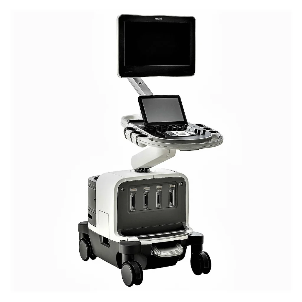
EPIQ Elite
Example of a premium multispecialty platform with probe families and functions suited to radiology, vascular, cardiac and OB/GYN.
View manufacturer’s overview ↗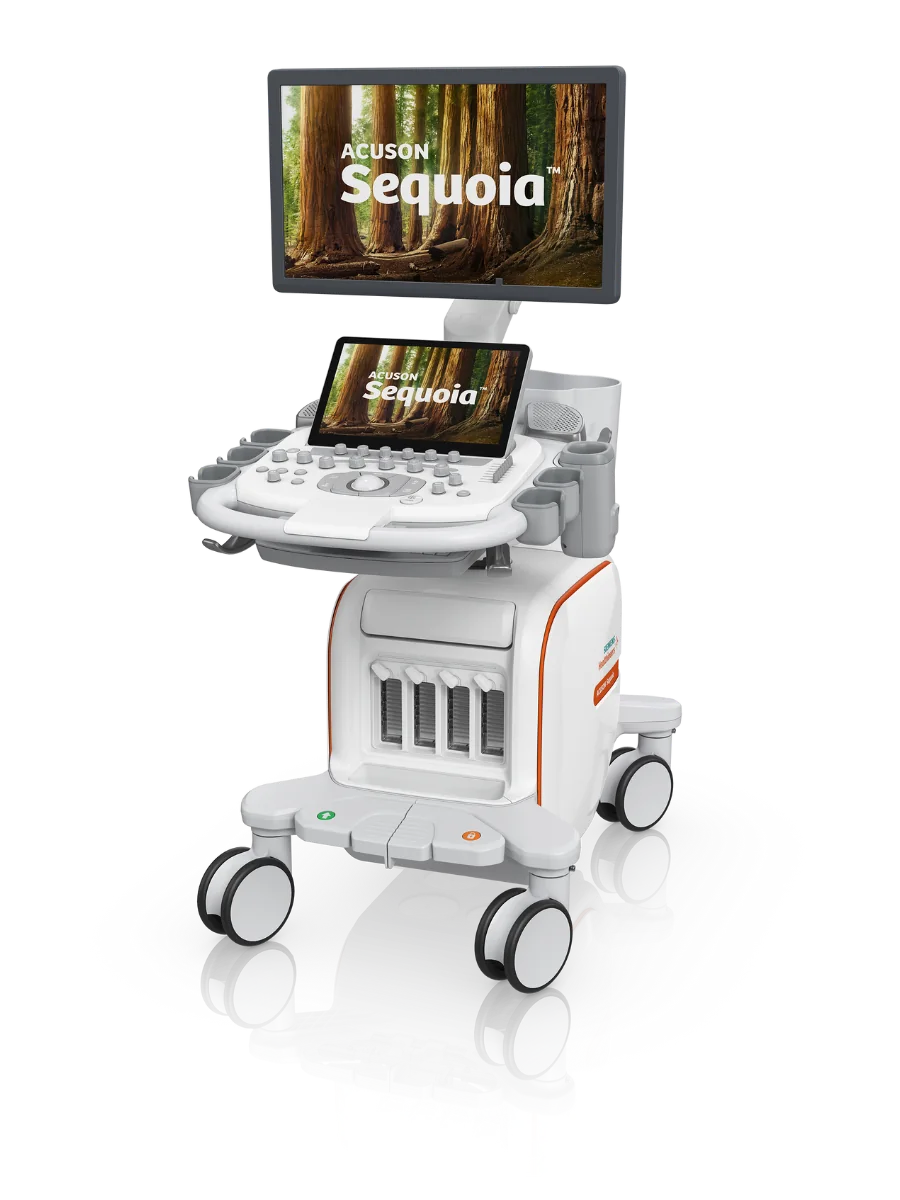
ACUSON Sequoia
Example of a shared-service system geared towards advanced imaging, notably for the deep abdomen, elastography, contrast and cardiovascular, depending on options.
View manufacturer’s overview ↗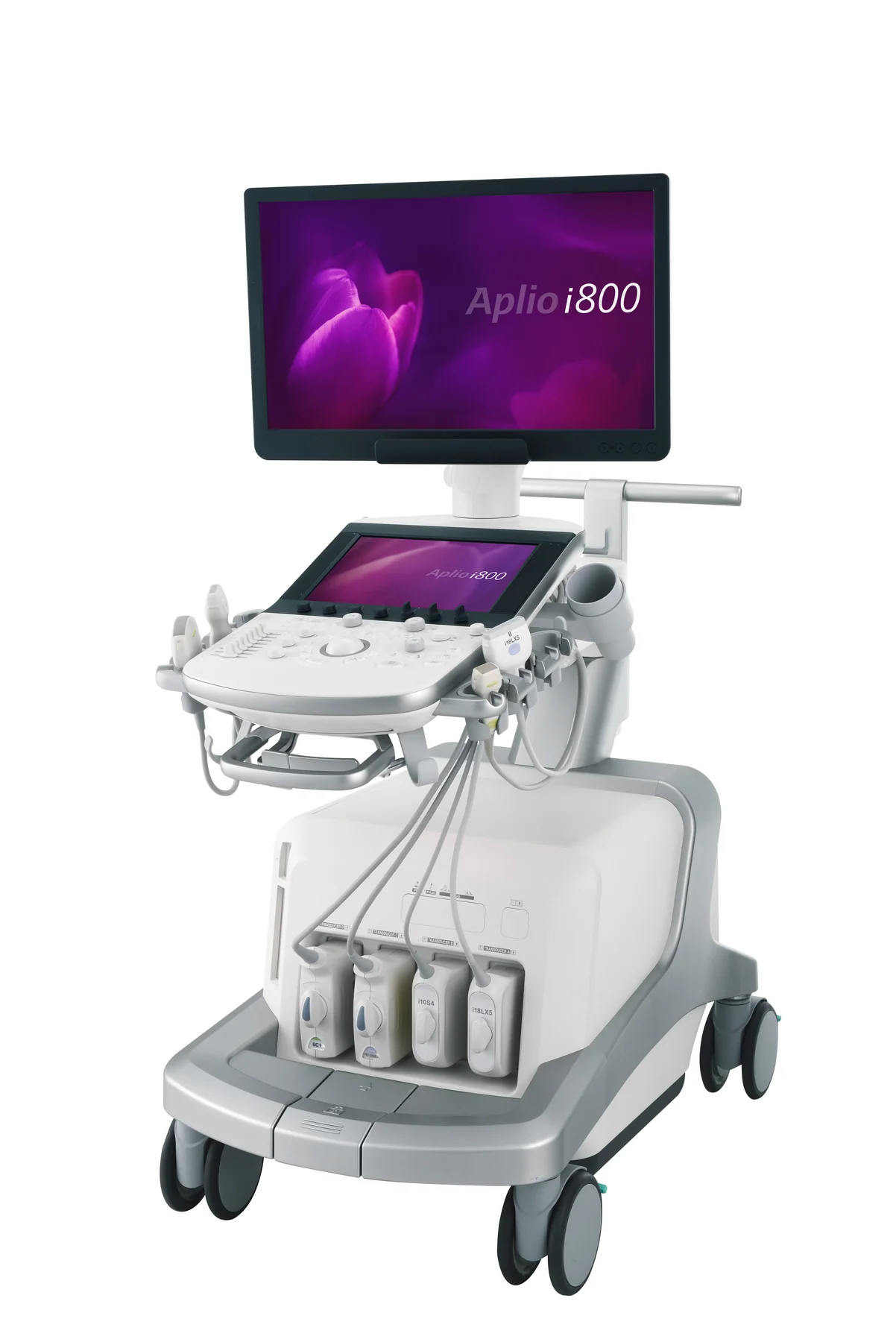
Aplio i-series
Example of a versatile platform combining general imaging, Doppler, microvascular imaging, elastography and interventional guidance, depending on licences.
View manufacturer’s overview ↗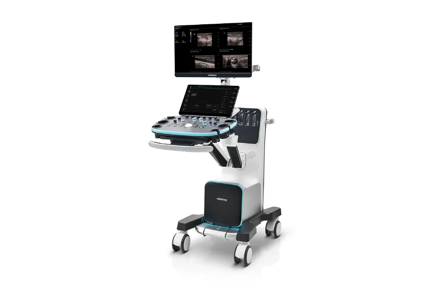
Resona I9
Example of a general-purpose system emphasising ergonomics, mobility, automation and an expandable set of clinical applications.
View manufacturer’s overview ↗Test with your users and your difficult cases
A convincing demonstration should use representative patients, examinations that are actually performed and the probes offered in the quotation. Have several operators assess penetration, resolution, Doppler, measurement reproducibility, examination time and ergonomics.
Prepare the room, the network and the data pathway
Ultrasound requires less building work than a CT scanner, but the environment remains decisive: stable power supply, network sockets, suitable air conditioning, circulation space, lighting, privacy, access to handwashing and protected probe storage.
Before delivery, validate the DICOM addresses, worklist, PACS, report formats, user accounts, backups, updates, remote access and cybersecurity policy. A function listed in the brochure is only useful if it is enabled, tested and integrated.
Match each probe to its reprocessing protocol
The level of cleaning or disinfection depends on how the probe is used and on patient contact. Endocavity probes and interventional procedures require higher precautions than external ultrasound on intact skin. The products used must remain compatible with the manufacturer’s instructions.
Regularly inspect the acoustic lens, housing, cable, connector and covers. A damaged probe can degrade the image, pose an electrical risk or promote contamination. Use the appropriate gel and sheaths, with traceability that complies with local procedures.
Ultrasound does not use ionising radiation, but acoustic exposure must remain justified and optimised. Trained users monitor, in particular, the thermal and mechanical indices according to the examination.
Budget for the probes as much as for the console
The cost of ownership includes the system, probes, software, accessories, network integration, training, updates, quality control, maintenance and downtime. Probes are among the components most exposed to impacts, twisting, fluid ingress and incompatible cleaning products.
Eight questions before signing
- Which applications and examinations will actually be performed from the first year?
- Which probes, licences, measurements and functions are included in the quoted price?
- Has image quality been assessed on our patients and our priority clinical cases?
- Does the system integrate with the network, DICOM, PACS, the worklist and cybersecurity requirements?
- What are the warranty terms for the console and for each probe?
- Who provides installation, applications training and maintenance, and what are the response times?
- Which cleaning products are compatible with the console and each probe type?
- What budget should be planned for probes, batteries, licences, carts, printers and accessories over five years?
See the technology in action
This video complements the guide with a visual introduction to ultrasound. It plays right here, without leaving Bio-MedX.
Reference points to adapt to the clinical context
Final requirements must be validated with the users, biomedical engineering, infection control, IT, the manufacturer and the competent authorities. The brands mentioned serve only as examples of platforms available on the market.